

鼠型斑疹伤寒立克次体IgG免疫荧光玻片
| 型 号: | |
| 报 价: |  |
鼠型斑疹伤寒立克次体IgG免疫荧光玻片 立克次体 巴尔通体 需要了解更多产品可以咨询我们,本产品由广州健仑生物科技有限公司提供
- 产品描述
鼠型斑疹伤寒立克次体IgG免疫荧光玻片
Rickettsia typhi IgG IFA Kit
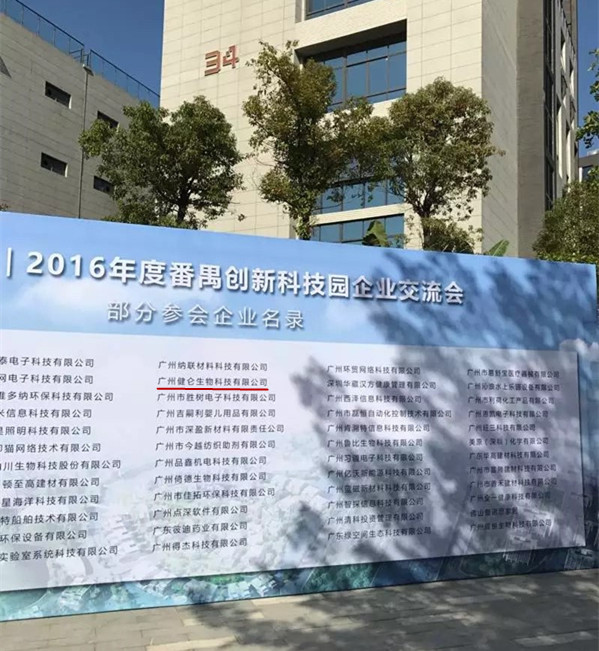
广州健仑生物科技有限公司
主要用途:用于检测人血清中的鼠型斑疹伤寒立克次体IgG抗体
产品规格:12 孔/张,10 张/盒
主要产品包括:包柔氏螺旋体菌、布鲁氏菌、贝纳特氏立克次体、土伦杆菌、钩端螺旋体、新型立克次体、恙虫病、立克次体、果氏巴贝西虫、马焦虫、牛焦虫、利什曼虫、新包虫、弓形虫、猫流感病毒、猫冠状病毒、猫疱疹病毒、犬瘟病毒、犬细小病毒等病原微生物的 IFA、MIF、ELISA试剂。
鼠型斑疹伤寒立克次体IgG免疫荧光玻片
我司还提供其它进口或国产试剂盒:登革热、疟疾、西尼罗河、立克次体、无形体、蜱虫、恙虫、利什曼原虫、RK39、汉坦病毒、深林脑炎、流感、A链球菌、合胞病毒、腮病毒、乙脑、寨卡、黄热病、基孔肯雅热、克锥虫病、违禁品滥用、肺炎球菌、军团菌、化妆品检测、食品安全检测等试剂盒以及日本生研细菌分型诊断血清、德国SiFin诊断血清、丹麦SSI诊断血清等产品。
欢迎咨询
欢迎咨询2042552662

| JL-FL31 | 康氏立克次氏体IgM ELISA | Rickettsia conorii IgM ELISA Kit | 用于检测人血清中的康氏立克次氏体IgM抗体 |
| JL-FL32 | 斑点热立克次体IgG ELISA | Spotted fever group IgG ELISA Kit | 用于检测人血清中的斑点热立克次体IgG抗体 |
| JL-FL33 | 斑疹伤寒立克次体IgG ELISA | Typhus group IgG ELISA Kit | 用于检测人血清中的斑疹伤寒立克次体IgG抗体 |
| JL-FL34 | 试剂盒 | Rickettsia typhi IgG IFA Kit | 用于检测人血清中的鼠型斑疹伤寒立克次体IgG抗体 |
| JL-FL35 | 鼠型斑疹伤寒立克次体IgG ELISA | R. typhi IgG ELISA Kit | 用于检测人血清中的鼠型斑疹伤寒立克次体IgG抗体 |
| JL-FL36 | 鼠型斑疹伤寒立克次体IgM ELISA | R. typhi IgM ELISA Kit | 用于检测人血清中的鼠型斑疹伤寒立克次体IgG抗体 |
| JL-FL37 | akari立克次体 IgG ELISA | R. akari IgG ELISA Kit | 用于检测人血清中的akari立克次体IgG抗体 |
| JL-FL38 | parkeri立克次体IgG ELISA | R. parkeri IgG ELISA Kit | 用于检测人血清中的parkeri立克次体IgG抗体 |
| JL-FL39 | montanensis立克次体IgG ELISA | R. montanensis IgG ELISA Kit | 用于检测人血清中的montanensis立克次体IgG抗体 |
| JL-FL40 | EB病毒衣壳IgG免疫荧光玻片试剂盒 | EBV Viral Capsid IgG IFA Kit | 用于检测人血清中的EB病毒衣壳IgG抗体 |
| JL-FL41 | EB病毒衣壳IgM免疫荧光玻片试剂盒 | EBV Viral Capsid IgM IFA Kit | 用于检测人血清中的EB病毒衣壳IgM抗体 |
| JL-FL42 | EB病毒早期抗原IgG免疫荧光玻片试剂盒 | EBV Early Antigens IgG IFA Kit | 用于检测人血清中的EB病毒早期抗原IgG抗体 |
二维码扫一扫
【公司名称】 广州健仑生物科技有限公司
【】 杨永汉
【】
【腾讯 】 2042552662
【公司地址】 广州清华科技园创新基地番禺石楼镇创启路63号二期2幢101-3室
【企业文化】

在所附上的视频上,显微照片的时间序列显示了,四杆状酵母细胞收缩环的形成和收缩。一收缩环的蛋白质显示为白色。该蛋白质簇环绕每个细胞的中间积累,然后冷凝成一个收缩环夹住细胞使之分为两个。
近期的研究结果证实了,新的心肌细胞可以低速率生成,表明了心脏干细胞的存在。但人们对于这些细胞的起源却并不清楚。
范德比尔特大学医学、细胞与发育生物学副教授Antonis Hatzopoulos博士和同事们推测,排列于血管上内皮细胞可能有潜力生成新的心肌细胞。他们知道,在发育过程中内皮细胞可生成包括血细胞在内的其他细胞类型。
现在,利用一些*技术在小鼠模型中“追踪”细胞,他们证实冠状动脉中的内皮细胞可以生成健康心脏中的新心肌细胞。他们发现在冠状动脉中有两种心脏干细胞群:静息的心脏干细胞群位于血管中膜层,增殖心脏干细胞群位于血管外膜层。
Hatzopoulos说,发现冠状动脉充当了心脏干细胞的“微环境”具有有趣的含义。美国的1号杀手:冠状动脉病会影响这一微环境。他说:“我们的研究表明,冠状动脉病不仅可以阻断动脉,引起心脏病发作,还可通过影响心脏维持和再生的途经来导致心力衰竭。”
在以往的研究中,Hatzopoulos和同事们证实心脏病发作后,内皮细胞可生成一些产生瘢痕组织的成纤维细胞。“看起来相同的内皮系统在稳态过程中生成了肌细胞,并在心肌梗死后转换生成了瘢痕组织。损伤之后,再生转向了纤维化,”他说。
了解这种转换有可能促成一些新的策略,在心脏病发作后、衰老过程中或在诸如糖尿病及高血压等疾病中再生和生成新的心肌。
Hatzopoulos说:“如果我们可以了解损伤后发生的这种命运转换的分子调控机制,或许我们就可以采用某种化合物或药物来恢复再生,生成肌肉而不是瘢痕。我们认为这是一次机会来改善我们对心肌梗死后来到诊所的患者的治疗方式。
近期的研究结果证实了,新的心肌细胞可以低速率生成,表明了心脏干细胞的存在。但人们对于这些细胞的起源却并不清楚。
范德比尔特大学医学、细胞与发育生物学副教授Antonis Hatzopoulos博士和同事们推测,排列于血管上内皮细胞可能有潜力生成新的心肌细胞。他们知道,在发育过程中内皮细胞可生成包括血细胞在内的其他细胞类型。
现在,利用一些*技术在小鼠模型中“追踪”细胞,他们证实冠状动脉中的内皮细胞可以生成健康心脏中的新心肌细胞。他们发现在冠状动脉中有两种心脏干细胞群:静息的心脏干细胞群位于血管中膜层,增殖心脏干细胞群位于血管外膜层。
Hatzopoulos说,发现冠状动脉充当了心脏干细胞的“微环境”具有有趣的含义。美国的1号杀手:冠状动脉病会影响这一微环境。他说:“我们的研究表明,冠状动脉病不仅可以阻断动脉,引起心脏病发作,还可通过影响心脏维持和再生的途经来导致心力衰竭。”
在以往的研究中,Hatzopoulos和同事们证实心脏病发作后,内皮细胞可生成一些产生瘢痕组织的成纤维细胞。“看起来相同的内皮系统在稳态过程中生成了肌细胞,并在心肌梗死后转换生成了瘢痕组织。损伤之后,再生转向了纤维化,”他说。
On the attached video, the time series of photomicrographs shows the formation and contraction of the collapsible rings of four-rod yeast cells. A shrunken protein appears white. The protein cluster accumulates around the middle of each cell, then condenses into a constriction ring that clamps the cell and breaks it into two.
Recent findings confirm that new cardiomyocytes can be generated at low rates, indicating the presence of cardiac stem cells. But people do not know the origin of these cells.
Antonis Hatzopoulos, Ph.D., associate professor of medicine, cell and developmental biology at Vanderbilt University, and colleagues speculate that lining up vascular endothelial cells may have the potential to generate new cardiomyocytes. They know that endothelial cells can generate other cell types, including blood cells, during development.
Now, using advanced techniques to "track" cells in mouse models, they demonstrate that endothelial cells in the coronary arteries produce new cardiomyocytes in healthy hearts. They found that there are two groups of cardiac stem cells in the coronary arteries: the resting cardiac stem cell population is located in the vascular media layer, and the proliferating cardiac stem cell population is located in the outer vascular layer.
Hatzopoulos said it was interesting to find that the coronary arteries act as a "microenvironment" for heart stem cells. No. 1 killer in the United States: Coronary artery disease can affect this microenvironment. "Our research shows that coronary artery disease can not only block the arteries and cause heart attacks but can also lead to heart failure through pathways that affect heart maintenance and regeneration," he said.
In previous studies, Hatzopoulos and colleagues confirmed that after a heart attack, endothelial cells produce some scar tissue-producing fibroblasts. "The seemingly identical endothelium generates muscle cells during homeostasis and converts to scar tissue after myocardial infarction, and after the injury, the regenerative turns to fibrosis," he said.
Understanding this transition may lead to new strategies to regenerate and generate new myocardium during a heart attack, during aging or in diseases such as diabetes and hypertension.
Hatzopoulos said: "If we can understand the molecular mechanisms of this fate shift that occur after a lesion, perhaps we could use a compound or drug to regain regeneration and produce muscle instead of scar, and we think this is an opportunity to improve us Treatment of patients who come to the clinic after myocardial infarction.
Recent findings confirm that new cardiomyocytes can be generated at low rates, indicating the presence of cardiac stem cells. But people do not know the origin of these cells.
Antonis Hatzopoulos, Ph.D., associate professor of medicine, cell and developmental biology at Vanderbilt University, and colleagues speculate that lining up vascular endothelial cells may have the potential to generate new cardiomyocytes. They know that endothelial cells can generate other cell types, including blood cells, during development.
Now, using advanced techniques to "track" cells in mouse models, they demonstrate that endothelial cells in the coronary arteries produce new cardiomyocytes in healthy hearts. They found that there are two groups of cardiac stem cells in the coronary arteries: the resting cardiac stem cell population is located in the vascular media layer, and the proliferating cardiac stem cell population is located in the outer vascular layer.
Hatzopoulos said it was interesting to find that the coronary arteries act as a "microenvironment" for heart stem cells. No. 1 killer in the United States: Coronary artery disease can affect this microenvironment. "Our research shows that coronary artery disease can not only block the arteries and cause heart attacks but can also lead to heart failure through pathways that affect heart maintenance and regeneration," he said.
In previous studies, Hatzopoulos and colleagues confirmed that after a heart attack, endothelial cells produce some scar tissue-producing fibroblasts. "The seemingly identical endothelium generates muscle cells during homeostasis and converts to scar tissue after myocardial infarction, and after the injury, the regenerative turns to fibrosis," he said.
