

钩端螺旋体IgG免疫荧光试剂盒
| 型 号: | |
| 报 价: |  |
钩端螺旋体IgG免疫荧光试剂盒 立克次体 巴尔通体 需要了解更多产品可以咨询我们,本产品由广州健仑生物科技有限公司提供
- 产品描述
钩端螺旋体IgG免疫荧光试剂盒
Leptospira IgG IFA Kit
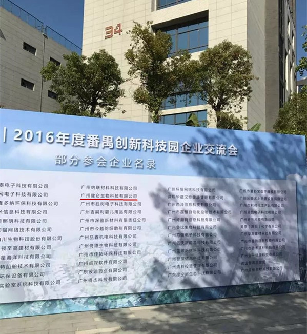
广州健仑生物科技有限公司
主要用途:用于检测人血清中的钩端螺旋体IgG抗体
产品规格:12 孔/张,10 张/盒
主要产品包括:包柔氏螺旋体菌、布鲁氏菌、贝纳特氏立克次体、土伦杆菌、钩端螺旋体、新型立克次体、恙虫病、立克次体、果氏巴贝西虫、马焦虫、牛焦虫、利什曼虫、新包虫、弓形虫、猫流感病毒、猫冠状病毒、猫疱疹病毒、犬瘟病毒、犬细小病毒等病原微生物的 IFA、MIF、ELISA试剂。
钩端螺旋体IgG免疫荧光试剂盒
我司还提供其它进口或国产试剂盒:登革热、疟疾、西尼罗河、立克次体、无形体、蜱虫、恙虫、利什曼原虫、RK39、汉坦病毒、深林脑炎、流感、A链球菌、合胞病毒、腮病毒、乙脑、寨卡、黄热病、基孔肯雅热、克锥虫病、违禁品滥用、肺炎球菌、军团菌、化妆品检测、食品安全检测等试剂盒以及日本生研细菌分型诊断血清、德国SiFin诊断血清、丹麦SSI诊断血清等产品。
欢迎咨询
欢迎咨询2042552662

| JL-FL38 | parkeri立克次体IgG ELISA | R. parkeri IgG ELISA Kit |
| JL-FL39 | montanensis立克次体IgG ELISA | R. montanensis IgG ELISA Kit |
| JL-FL40 | EB病毒衣壳IgG免疫荧光玻片试剂盒 | EBV Viral Capsid IgG IFA Kit |
| JL-FL41 | EB病毒衣壳IgM免疫荧光玻片试剂盒 | EBV Viral Capsid IgM IFA Kit |
| JL-FL42 | EB病毒早期抗原IgG免疫荧光玻片试剂盒 | EBV Early Antigens IgG IFA Kit |
| JL-FL43 | Leptospira IgG IFA Kit | |
| JL-FL44 | 钩端螺旋体IgM免疫荧光试剂盒 | Leptospira IgM IFA Kit |
| JL-FL45 | 果氏巴贝西虫免疫荧光玻片 | Babesia microti IFA Substrate slide |
| JL-FL46 | 果氏巴贝西虫IgG免疫荧光试剂盒 | Babesia microti IgG IFA Kit |
| JL-FL47 | 果氏巴贝西虫IgM免疫荧光试剂盒 | Babesia microti IgM IFA Kit |
| JL-FL48 | 埃立克体IgG微量免疫荧光试剂盒 | Ehrlichia canis Canine IFA IgG Kit |
| JL-FL49 | 包柔氏螺旋体菌IgG免疫荧光试剂盒 | Borrelia IgG IFA Kit |
| JL-FL50 | 布鲁氏菌IgG免疫荧光试剂盒 | Brucella IgG IFA Kit |
| JL-FL51 | 里氏新立克次体IgG免疫荧光试剂盒 | Neorickettsia risticii IgG IFA Kit |
| JL-FL52 | 弓形虫IgG免疫荧光试剂盒(检测猫) | Toxoplasma IFA Feline IgG Kit |
| JL-FL53 | 弓形虫IgG免疫荧光试剂盒(检测狗) | Toxoplasma IFA Canine IgG Kit |
二维码扫一扫
【公司名称】 广州健仑生物科技有限公司
【】 杨永汉
【】
【腾讯 】 2042552662
【公司地址】 广州清华科技园创新基地番禺石楼镇创启路63号二期2幢101-3室
【企业文化】

当这些细胞球体在悬浮培养基中继续保存了数天之后,视网膜上皮组织自发地向外突出,形成视泡样的结构。然后,突起部分的顶端又开始内陷,形成酒杯状结构,很像胚胎上眼睛的视杯。与活体动物中一样,这个源自胚胎干细胞的视杯也由内外两层组成:外壁是上皮层,内壁便是视网膜。换句话说,在培养皿中,原本分开的干细胞组成的聚集体,独自形成了有序的结构——真是名副其实的“eyepopping”(这个词有双关含义,从字面上理解,就是“眼睛出现”,而这个词本身的含义是“使人瞠目的”)。不像在胚胎中,培养皿中并没有晶状体和角膜。这一发现清楚地回答了那个存在已久的问题:原始视网膜的形成,是否需要晶状体等邻近组织施加外部作用力。至少在体外,视网膜的形成是一个基于细胞内部程序的自发现象。
眼睛的形成
由一小块胚胎组织发育成眼睛,会经历如下步骤:内部的神经上皮层向外凸起,形成视泡(第9天);凸起部分的外层向内凹陷(第9.5天),晶状体泡随之形成(第10天);视泡的一部分发生折叠,形成视杯,与晶状体泡共同形成视网膜、视神经及zui外部的晶状体(第10.5天)。视网膜包括三个不同的细胞层:视杆和视锥细胞;水平、双极和无长突细胞;视神经节细胞。
培养皿中,眼睛的发育过程继续进行着,就像我们在胚胎发育中所看到的一样。我们让视杯在悬浮培养体系中又呆了两个星期,组织的直径大约长到了2毫米,而且与胚胎中一样,单层的视网膜上皮也演变成了层级结构,含有所有6种可在初生婴儿的眼睛中检测到的细胞。
这一层级结构的外层是光感受器细胞层,zui内层是神经节细胞,这类细胞在机体中的作用是连接视网膜与大脑。如同你在真实视网膜中看到的一样,在内外两层之间是几层连接层,由中间神经元(interneuron)构成。与此前一样,多层结构的出现也是依照内在程序完成,这个程序会决定什么细胞应该产生,它们又应该被安排在三维空间的什么位置。
我们的工作仍未结束。视杯究竟如何形成,一团细胞究竟如何出现规整的结构,这些问题仍然存在。由同一物质构成的团块自发产生复杂形状,这一过程叫做对称破缺(symmetry breaking),贯穿整个胚胎发育过程。如果没有对称破缺,受精卵重复进行细胞分裂只会产生一团未分化的细胞,发育过程会止步不前。我们的自组织胚胎干细胞培养体系似乎可以作为一个理想的实验平台,来研究哺乳动物胚胎形成过程中的这些玄妙机制。
Retinal epithelial tissue spontaneously protrudes outwardly to form a retinoid-like structure when these cell spheres are kept in suspension for several days. Then, the top of the protuberance begins to invaginate, forming a goblet-like structure much like the optic cup of the eye on the embryo. Like in live animals, this embryonic stem cell-derived optic cup is also composed of two layers: the outer wall is the epithelium and the inner wall is the retina. In other words, in petri dishes, the aggregates of originally separate stem cells form an ordered structure alone - a veritable "eyepopping" (the word has the double meaning, literally, "eyes appear" , And the word itself means "deception"). Unlike in embryos, there are no lenses and corneas in the petri dish. This finding clearly answers the long-standing question of whether the formation of the primordial retina requires the application of external forces to nearby tissues such as the lens. Retinal formation, at least in vitro, is a spontaneous phenomenon based on intracellular processes.
Eye formation
The development of the eye from a small piece of embryonic tissue proceeds through the following steps: The inner neuroepithelial layer bulges outward to form a retinal bulb (day 9); the outer layer of the convex portion is inwardly depressed (day 9.5), the lens The bubble then forms (Day 10); part of the optic disc folds to form a optic cup, which together with the lensblack forms the retina, the optic nerve, and the outermost lens (Day 10.5). The retina consists of three distinct layers of cells: rods and cones; horizontal, bipolar and amacrine cells; optic ganglion cells.
Petri dishes, the development of the eye continued, as we have seen in embryonic development. We left the optic cup for another two weeks in the suspension culture system, and the tissue grew to about 2 mm in diameter, and as in the embryo, the monolayer of the retinal epithelium also evolved into a hierarchical structure containing all six Cells detected in the baby's eyes.
The outer layer of this hierarchy is the photoreceptor cell layer, the innermost layer is the ganglion cells, the role of these cells in the body is to connect the retina and the brain. As you can see in the real retina, there are several tie layers between the inner and outer layers, made up of interneurons. As before, the advent of multilayered structures is also done in accordance with an internal program that determines what cells should be produced and where they should be arranged in three dimensions.
Our work is still not over. How the formation of optician exactly how a group of cells appear structured structure, these problems still exist. Complex masses of spontaneously formed clumps of the same substance, called symmetry breaking, run through the entire embryonic development. If there is no symmetry breaking, repeated fertilization of eggs will only produce a cell division of undifferentiated cells, the development process will be halted. Our self-organizing embryonic stem cell culture system seems to be an ideal experimental platform to study these mysterious mechanisms in mammalian embryogenesis.
