

肠道感染诺如病毒快检卡(胶体金法)
| 型 号: | 西班牙certest |
| 报 价: |  |
肠道感染诺如病毒快检卡(胶体金法): 需要了解更多产品可以咨询我们,本产品由广州健仑生物科技有限公司提供
- 产品描述
肠道感染诺如病毒快检卡(胶体金法)
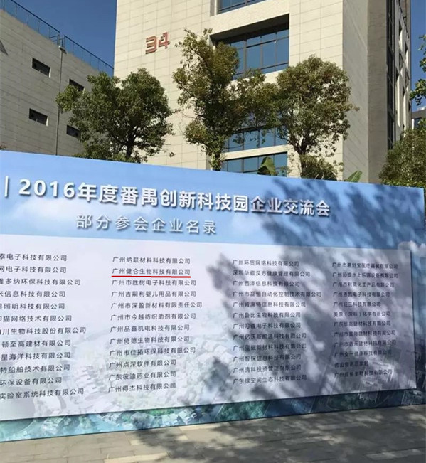
广州健仑生物科技有限公司
(广州健仑生物科技有限公司是集研制开发、销售、服务于一体的优良企业,公司产品涉及临床快速诊断试剂、食品安全检测试剂,违禁品快速检测,动物疾病防疫检测试剂,免疫诊断试剂、临床血液学和体液学检验试剂、微生物检验试剂、分子生物学检验试剂、临床生化试剂、有机试剂等众多领域,同时核心代理Panbio、FOCUS、Qiagen、IBL、CORTEZ、Fuller、Inbios、BinaxNOW、LumuQuick、日本富士、日本生研等多家有名诊断产品集团公司产品,致力于为商检单位、疾病预防控制中心、海关出入境检疫局、卫生防疫单位,缉毒系统,戒毒中心,检验检疫单位、生化企业、科研院所、医疗机构等机构与行业提供*、高品质的产品服务。此外,本公司还开展食品、卫生、环境、药品等多方面的第三方检测服务。)
主要用途:用于检测粪便标本中的诺如病毒抗原,以支持诺如病毒感染的诊断。
产品规格:20T/盒
存储条件:2-30℃
肠道感染诺如病毒快检卡(胶体金法)
我司还提供其它进口或国产试剂盒:登革热、疟疾、西尼罗河、立克次体、无形体、蜱虫、恙虫、利什曼原虫、RK39、汉坦病毒、深林脑炎、流感、A链球菌、合胞病毒、腮病毒、乙脑、寨卡、黄热病、基孔肯雅热、克锥虫病、违禁品滥用、肺炎球菌、军团菌、化妆品检测、食品安全检测等试剂盒以及日本生研细菌分型诊断血清、德国SiFin诊断血清、丹麦SSI诊断血清等产品。
欢迎咨询
欢迎咨询2042552662
【产品介绍】
二维码扫一扫
【公司名称】 广州健仑生物科技有限公司
【】 杨永汉
【】
【腾讯 】 2042552662
【公司地址】 广州清华科技园创新基地番禺石楼镇创启路63号二期2幢101-3室
【企业文化】
颈段上部近枕骨大孔处近似圆形,往下为 三角形,矢径短,横径长;胸段大致呈圆形;腰段上、中部呈三角形 ,下部呈三叶形;骶段呈扁三角形。椎管以第4~6胸椎zui为狭小,颈 段以第7颈椎、腰段以第4腰椎较小。先天性椎管狭窄是由于在脊柱的 生长形成中,包括营养外伤等因素造成椎管发育的先天性狭窄致病。 大部分患者开始无症状,到中年后由于脊柱的一些退行性病变或损伤 ,从而导致椎管狭窄的症状及体征出现。后天性椎管狭窄是由于椎间 盘突出、椎体增生、椎体滑脱以及后纵韧带、黄韧带增生肥厚、钙化 或骨化等刺激脊髓神经及周围血管,造成神管发生炎症粘连、充血、 水肿,从而导致椎管狭窄的发生神经管(neural tube)神经系统的 组成部分。起源于胚体背侧的神经外胚层。人胚第三周初,在脊索的 诱导下,胚体背侧外胚层加厚成一条纵长的神经板,随着脊索的延伸 ,神经板也在胚的纵轴上延伸。神经板由于周边部分生长较快,沿着 中轴下陷,形成神经沟,左右外缘弯向背侧隆起,形成左右神经褶。 随着神经沟的不断加深,两侧的神经则走子褶走越长越高,接近正中 线。在相当于枕部体节的平面上,由神经沟愈合而成。愈合过程向头 、尾两端进展,zui后在头尾两端各有一开口,分别称前神经孔和后神 经孔。胚胎第25天左右,前神经孔闭合,第27天左右,后神经孔闭合 ,形成完整的神经管。其前段膨大,衍化为脑;后段较细,衍化为脊 髓。形成的两种方式神经管是中枢神经系统的原基,其形成有两种方 式。1.Primary neurulation由外胚层细胞增殖、内陷、并zui终离开 外胚层表面而形成中空的神经管,大多数脊椎动物头部神经管采此种 方式它可分为三个过程:(1)Neuralplate(神经板)的形成:背部 mesoderm诱导中线外胚层细胞变长,而其侧翼的预定外胚层细胞变扁 平,使预定的神经区域凸出于周边外胚层而成为神经板;预定的表皮 细胞和神经细胞的运动导致二者交界处形成neuralfold(神经褶)。
The upper cervical segment near the foramen magnum is approximay circular, down to the triangle, short radius, long horizontal; thoracic segment is generally circular; waist segment, the middle triangular, trilobal lower; sacral segment was triangular. The 4th to 6th thoracic vertebrae were the most narrow, the cervical segment was the 7th cervical vertebra, and the lumbar segment was the 4th lumbar. Congenital spinal stenosis is due to the growth of the spine in the formation, including nutritional traumatic factors such as spinal canal congenital stenosis caused by disease. Most patients start asymptomatic, to middle-aged due to some of the spinal degenerative lesions or injuries, leading to spinal stenosis symptoms and signs appear. Acquired spinal canal stenosis is due to disc herniation, vertebral hyperplasia, vertebral body spondylolisthesis and posterior longitudinal ligament, ligamentum flavum hyperplasia hypertrophy, calcification or ossification and other spinal nerve and peripheral blood vessels, resulting in inflammation of the optic nerve adhesion, congestion, edema, As a result, spinal stenosis occurs as a component of the neural tube's nervous system. Originated from the back of the embryoid body neuroectoderm. At the beginning of the third week of the human embryo, under the guidance of the chordate, the dorsal ectoderm of the embryo body is thickened into an elongate nerve plate. Along with the extension of the notochord, the nerve plate also extends on the longitudinal axis of the embryo. Nerve plate due to the rapid growth of the peripheral part of the sunken along the axis, the formation of nerve ditch, the left and right outer edge curved dorsiflexion, the formation of left and right nerve fold. With the continuous deepening of the nerve groove, the nerves on both sides of the fold is walking taller and taller, close to the midline. Equivalent to the occipital section of the plane, from the nerve groove healing. Healing process to the head and tail both ends of the progress, and finally in both ends of the head and tail have an opening, respectively, before and after the nerve hole called the nerve hole. Around the 25th day of embryo, the anterior neural pore was closed and the 27th day after the closure of the neural pore, forming a complete neural tube. Its anterior enlargement, evolved into the brain; later smaller, evolved into the spinal cord. The two forms of neural tube is the central nervous system primordia, which formed in two ways. 1.Primary neurulation by ectodermal cells proliferation, invagination, and eventually leave the surface of the ectoderm to form a hollow tube, the majority of vertebrate head nerve canal in this way it can be divided into three processes: (1) Neuralplate (Neural plate) formation: the mesoderm on the back induces a lengthening of the cells of the midline ectoderm, while the flanking predetermined ectoderm cells become flattened so that the predetermined neural area protrudes from the peripheral ectoderm and becomes a nerve plate; the predetermined epidermal cells and nerves The movement of cells leads to the formation of neuralfold (nerve folds) at the junction of the two.
